

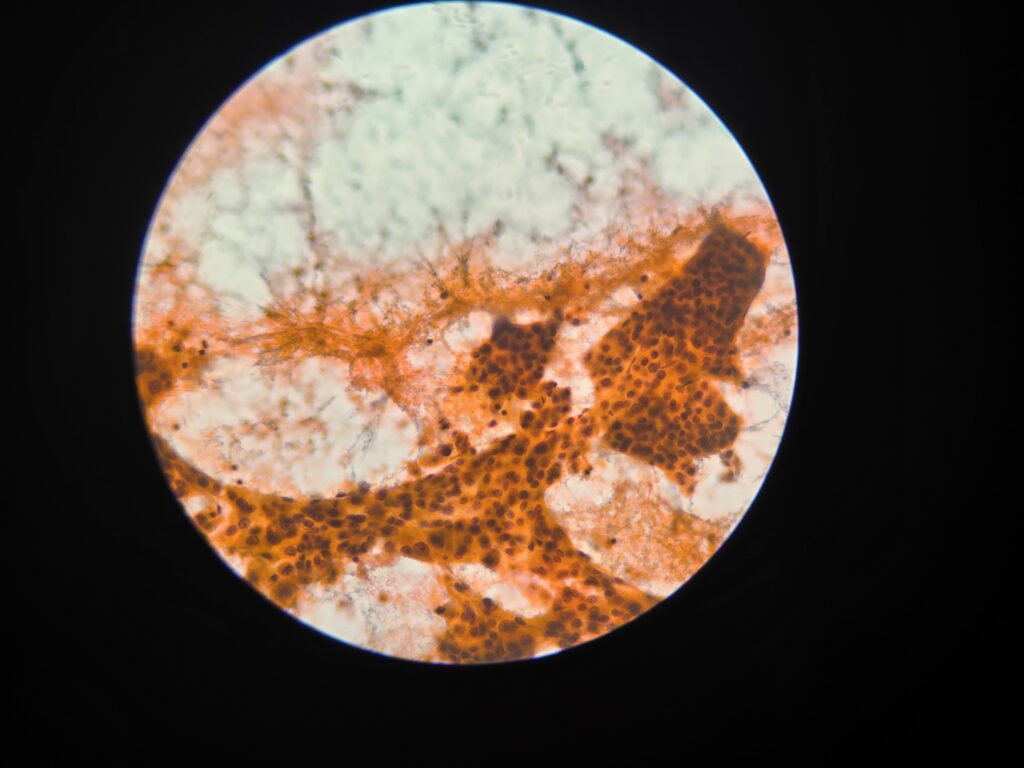
Hepatocellular carcinoma
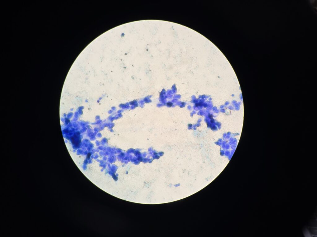
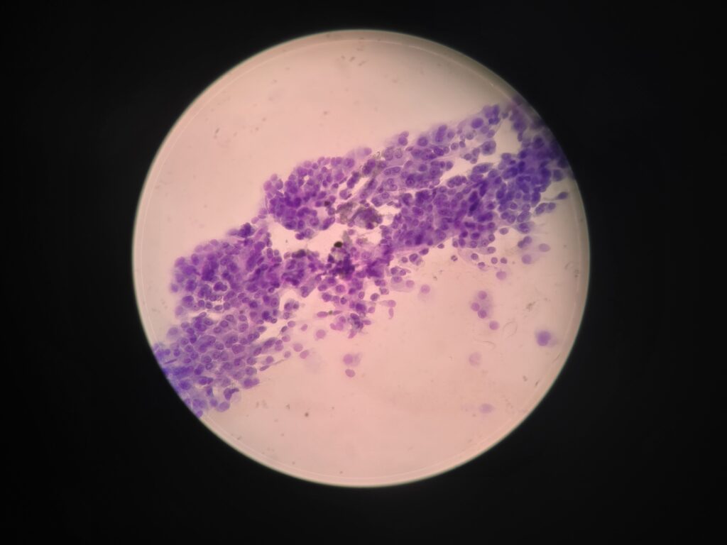
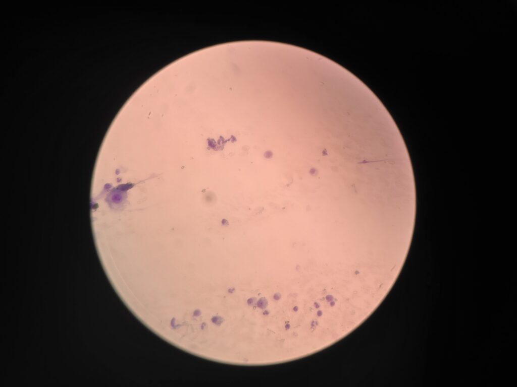
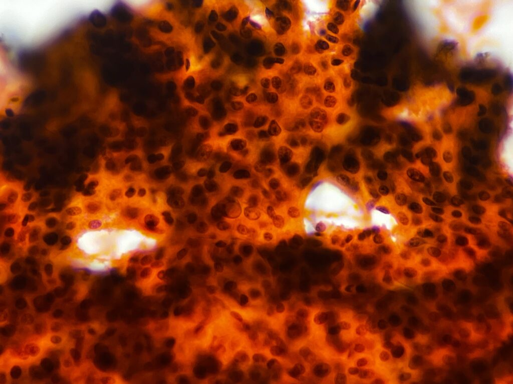
Pics 1 to 4 show clusters of cells in HCC on ROSE.
Pic 5 shows the trabecular pattern of arrangement of the hepatocytes (as seen in conventional HCC)
Pic 6 shows the endothelial cells lining the sinusoids. Normally, the hepatocytes are in linear pattern radiating from the portal vein to the central vein. They are usually 1 layer thick(sometimes 2 layers) but in a malignancy,they become more than 2 layered causing a widening of the trabeculae.
Pic 7 shows pseudoinclusion(right around the centre) along with endothelial cells.
CRITERIA FOR DIAGNOSIS
Cellular smears.
Neoplastic cells forming widened trabeculae, acini or multilayered sheets, Endothelial cells lining cell groupings or small capillaries transgressing clusters of tumor cells.
Polygonal cells with increased N:C ratio, Central round nuclei often with macronucleoli.Increased nuclear size and atypia with binucleation and multinucleation.
Scant to abundant eosinophilic, granular cytoplasm; may be vacuolated.Dispersed cells including many atypical stripped nuclei, Intranuclear cytoplasmic inclusions.
Intracytoplasmic eosinophilic fibrillary inclusions (hyaline bodies)Intracytoplasmic bile pigment and/or bile plugs between cells, green or yellow in Papanicolaou smears and greenish-black with MGG.Frequent mitoses Bile duct epithelium absent.Loss of the above characteristics with dedifferentiation.
The characteristic findings relate to) (1) cell groupings (2) relationship to endothelium and (3) morphology.The features in any individual FNA sample are extremely variable and largely dictated by tumor differentiation.As the degree of differentiation decreases,the cells become more obviously malignant and their resemblance to hepatocytes decreases.Smears are typically cellular with large fragments, clusters and(dispersed cells. Cell groupings are classically trabecular, particularly in better-differentiated tumors, Acinar arrangements may be seen in up to 40% of HCC. With decreasing differentiation, smaller sheets and single-lying cells become more frequent, A reticulin stain on smears or cell block material may highlight the widened trabeculae and/or acinar structures or the reduced or absent reticulin.Endothelial relationships to HCC cell groups are an integral part of the diagnosis.Endothelial cells of sinusoidal capillaries may traverse or enclose trabeculae or separate tumor cell groupings.This important diagnostic criterion is diminished and then lost with decreasing differentiation.The cells resemble hepatocytes, thus demonstrating polygonal outlines and dense, granular cytoplasm, but with increased nucleocytoplasmic ratios over normal counterparts. Centrally placed nuclei, characterized by granular to coarse chromatin, show cytoplasmic invaginations (intranuclear inclusions) in up to 70% of cases and large central nucleoli in up to 60% of cases.One of the most outstanding cytological appearances in HCC is the presence of many stripped malignant nuclei with identical characteristics, lying free in the background between cell groups.The presence among malignant cells of occasional tumor giant cells or of bizarre hepatocytes would seem to favor primary liver carcinoma as they are rare in metastatic carcinoma.Bile pigment within the cytoplasm and between the cells proves the hepatocellular origin of the tumor, but is demonstrable in only 25% of cases.Fouchet’s reagent counterstained with hematoxylin/Sirius red stains bile intensely green.
Source for text: Orell and Sterrett’s Fine Needle Aspiration Cytology, 5th edition.